In a significant leap for neuroscience, ultra-fine neural probes known as Neuropixels are now being used to record neuronal activity in the human brain. This cutting-edge technology, detailed in a recent Nature report, allows researchers to observe brain function at the single-neuron level, offering unprecedented insights into how the brain processes information.
A Microscopic Window into the Brain
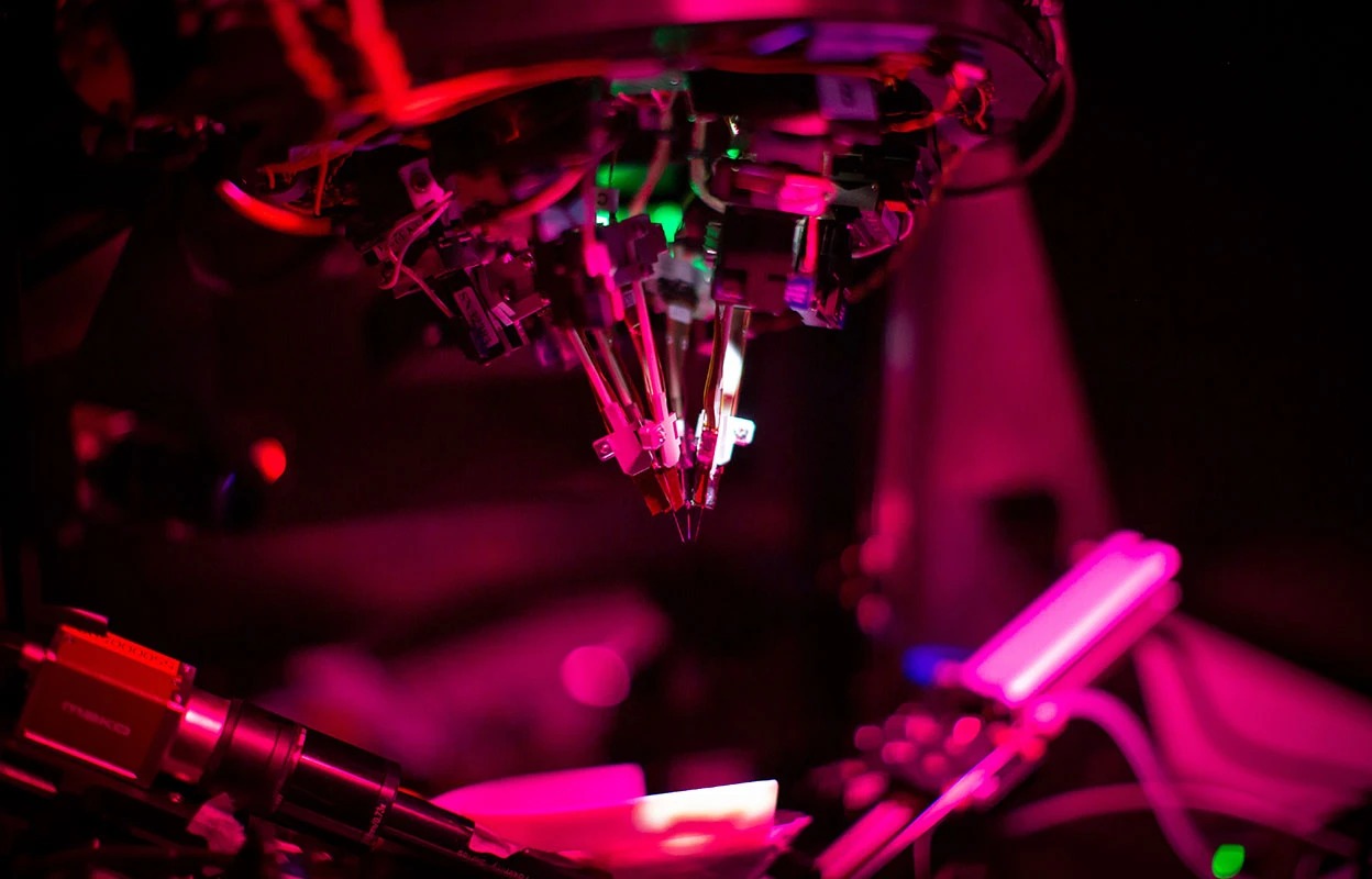
Neuropixels probes, developed through a collaboration involving the Howard Hughes Medical Institute’s Janelia Research Campus and Imec, are silicon-based devices designed for high-density recording of neuronal electrical activity. These probes are remarkably thin, measuring about 1 centimeter in length and narrower than a human hair. Their advanced design enables them to simultaneously record the electrical activity of hundreds, and even thousands, of neurons.
Unlike traditional electroencephalography (EEG) methods that capture collective signals from large groups of neurons, Neuropixels provide a much more granular view. They allow researchers to monitor individual neuron activity, offering finer data on how different brain regions process and transmit information. Pierre Bourdillon, a neurosurgeon at Fondation Rothschild Hospital in Paris, likened the technology’s impact to equipping every individual in a crowd with a personal microphone, a stark contrast to the previous method of placing a single microphone in a busy intersection.
First Human Trials Record Language Response
In a groundbreaking experiment conducted at the National Hospital for Neurology and Neurosurgery in London, researchers utilized the Neuropixels probe with a female patient undergoing surgery for normal pressure hydrocephalus. Neurosurgeon Ahmed Toma briefly inserted the probe into the patient’s brain during the procedure, while researchers from the Francis Crick Institute monitored the neural signals in real-time.
During the trial, the patient listened to a series of repeated words, such as “blind spot,” “parking spot,” and “piggy bank.” The objective was to observe how the brain represents transitions between different stimuli and how factors like word frequency and familiarity influence neural activity. The monitoring screens displayed clear changes in neural signal waveforms as the sounds played, demonstrating the probe’s capability to capture the human brain’s real-time response to linguistic stimuli.
Evolution of Neuropixels Technology
First introduced in 2017, the initial generation of Neuropixels probes featured 960 recording sites on their silicon bodies, with 384 capable of simultaneous signal reading. These probes were connected to a small chip, about the size of a Nano SIM card, responsible for reading, amplifying, and digitizing neural signals.
Early experiments using the first-generation probes involved implanting two probes into the brains of mice, simultaneously recording the activity of approximately 700 neurons across five brain regions. The subsequent Neuropixels 2.0, released in 2021, further refined the technology by reducing probe size and weight, while increasing the number of recording sites to 5120, with 1536 capable of simultaneous reading.
These advancements have enabled researchers to record the activity of tens of thousands of neurons simultaneously in animal models. Studies have extended to track neuronal activity for months and have broadened to encompass multiple brain regions, facilitating the analysis of how different areas collaborate in processes like sensation, movement, cognition, and decision-making.
A large-scale collaborative study by the International Brain Laboratory, involving 12 labs, is set to utilize 699 Neuropixels probes in 2025. This ambitious project aims to record neural activity from 139 mice across 279 brain regions, generating data from over 600,000 neural activity units. The goal is to create a comprehensive neural activity atlas mapping processes such as visual stimuli, movement, and decision-making.
Widespread Adoption and Future Prospects
Currently, approximately 1,700 laboratories worldwide are employing Neuropixels technology. The probes have been used to study a diverse range of over 20 animal species, including zebrafinches, bearded dragons, and octopuses, as well as non-human primates.
Tirin Moore, a neuroscientist at Stanford University, highlighted the dramatic increase in data collection capabilities. Before Neuropixels, researchers could typically record the activity of only about 20 non-human primate neurons per day, even in successful experiments. Now, his team routinely records the activity of at least 100 neurons. Moore and his colleagues plan to use these probes to map the visual system of macaques in high resolution, aiming to deepen the understanding of visual information processing in the brain.
A Tool for Discovery, Not Yet Clinical Therapy
It is crucial to note that Neuropixels probes are primarily utilized for fundamental neuroscience research, aiding scientists in understanding brain information processing mechanisms. They are not yet mature brain-computer interfaces or clinical therapeutic devices.
While this research holds immense promise for future advancements in brain-computer interface technology and the study of neurological disorders, it should not be mistaken for established clinical treatments. The progression from mouse models to non-human primates and now to brief recordings during human surgery marks a significant step towards single-cell level analysis of neural activity. Neuropixels are empowering researchers to not only observe brain region activity but also to dissect the role of individual neurons in complex cognitive processes.